If your full-arch implant cases still require verification jigs, multiple appointments, or last-minute adjustments, your workflow is costing you time, money, and predictability.
Understanding what photogrammetry is (and what it replaces) starts with an honest look at where current methods still fall short.
For years, clinicians relied on analog techniques like lost wax casting, where shrinkage and distortion made passive fit difficult to achieve. Even today, standard intraoral scanners can lose tracking across wide, edentulous spans, introducing stitching errors that compromise accuracy.
With accuracy as precise as 5 microns, photogrammetry allows dentists to eliminate verification jigs, significantly reduce chair time, and achieve a truly passive fit in full-arch cases using multi-unit abutments (MUAs).
- What Is Dental Photogrammetry and Why Does It Matter?
- Photogrammetry vs. Traditional Intraoral Scanning: What’s the Difference?
- Step-by-Step: How Does the Photogrammetry Workflow Operate?
- What Are the Benefits of Photogrammetry in Full-Arch Implants?
- In-House Photogrammetry vs. Outsourcing: Should You Partner With a Lab?
What Is Dental Photogrammetry and Why Does It Matter?
Photogrammetry is a technique that combines photography, mathematics, and computer science to generate highly accurate 3D models from 2D images.
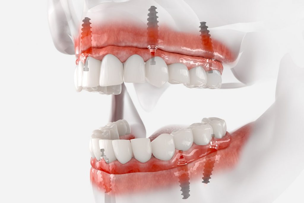
In implant dentistry, it is used to capture the precise positions and angulations of dental implants in three dimensions.
Unlike traditional impressions or intraoral scans alone, photogrammetry focuses on one thing with extreme precision: where the implants actually are in space.
According to a study published in the Dentistry Journal, photogrammetry produced digital dental models with high precision and trueness, supporting its potential as an alternative to conventional scanning for certain applications.
Unlike traditional impressions, photogrammetry uses a non-contact sensor system, which means the patient has greater comfort and less restriction during the scanning process.
Why does that precision matter? As Shawn Parisien, Laboratory Manager at Watersedge Dental Laboratory, explains:
If a structure doesn’t fit passively, but it is screwed down onto the implants, the implants receive the tension of that malfitting structure… The bone doesn’t like that. It lends itself over time to implant failure.
Photogrammetry vs. Traditional Intraoral Scanning: What’s the Difference?

Traditional intraoral scanners function as a wand inside the mouth, capturing detailed images of soft-tissue geometry and tooth contours.
In contrast, photogrammetry operates extraorally (from outside the oral cavity). The scanner only identifies specific physical reference points and does not scan the patient’s gums, tongue, or facial structure.
As Parisien puts it:
It doesn’t look for soft tissue, doesn’t look for tongue, doesn’t look for face, doesn’t look for implants. It looks for dominoes.
Those “dominoes” are the small, domino-shaped scan bodies screwed temporarily onto each MUA during the scan.
To create a complete picture, a small titanium fiduciary marker is placed in the mouth (typically on the palate), recognized by both the photogrammetry scan and the intraoral scan, allowing software to merge the two datasets seamlessly.
The result is a comprehensive digital model that integrates precise implant coordinates with the patient’s soft-tissue anatomy.
It is important to note that photogrammetry does not replace intraoral scanning. The two technologies work together, each fulfilling a distinct role.
Step-by-Step: How Does the Photogrammetry Workflow Operate?
1. Image Capture & Reference Points
The process begins by attaching specialized temporary scan bodies to the MUAs. A camera system such as our Imetric ICam4D captures multiple overlapping high-resolution photographs from various angles.
2. Feature Matching & Data Processing
Software algorithms use computer vision and triangulation to identify common features and reference points across the overlapping images, calculating the spatial relationship between each implant.
3. 3D Model Creation
The software processes this data into a precise 3D map of the implant positions. This information flows directly into dental planning software to record the exact angle and placement of each implant, then merges with the intraoral scan using the fiduciary marker.
What Are the Benefits of Photogrammetry in Full-Arch Implants?
Unmatched Precision & Passive Fit
While traditional analog workflows can leave up to 100 microns of discrepancy between implants, photogrammetry systems like Imetric tighten that to just 5 microns.
In fact, a systematic review and meta-analysis found that stereophotogrammetry demonstrated higher precision than intraoral scanning for complete-arch implant impressions, supporting its use in fabricating full-arch prostheses.
This level of accuracy ensures a passive-fit prosthesis that minimizes stress and strain on the supporting implants. It is precise enough that some manufacturers guarantee the fit of substructures fabricated from photogrammetry scans.
Fewer Appointments & Shorter Chair Time
Since the digital models are so accurate, photogrammetry can eliminate the traditional verification jig appointment entirely. Parisien estimates that most cases eliminate approximately three appointments compared to the traditional workflow.
For clinicians whose overhead runs around $475 per hour of chair time, the savings are significant. Patients benefit from fewer appointments, less time away from work, and a faster path to final restoration.
Broader Dental Applications
Beyond implant workflows, related forms of photogrammetry and stereophotogrammetry have also been explored in facial analysis and other digital dental applications. However, the strongest current evidence supports its use in full-arch implant impression accuracy.
In-House Photogrammetry vs. Outsourcing: Should You Partner With a Lab?
Purchasing advanced photogrammetry equipment requires a significant financial investment. Parisien shares that it’s “approaching six figures in total investment costs”, including hardware, software, and training.
Beyond cost, there is a steep learning curve to master the specialized software and data integration processes. For a practice that only handles a handful of full-arch cases per year, the investment is difficult to justify.
By partnering with a fully equipped dental lab like Watersedge, practices can access industry-leading scanners, expert digital workflows, and trained technicians without taking on the overhead.
Conclusion: Bringing High-Tech Precision to Your Chairside
Integrating photogrammetry into your practice offers undeniable clinical benefits: faster, cleaner immediate load deliveries at the surgical stage and highly predictable, stress-free final restorations.
Ottawa-area dentists do not need to take on the heavy overhead. Watersedge Dental Laboratory has already built a mature digital ecosystem and absorbed the investment.
Working with Watersedge gives your clinic access to the Imetric ICam system and our highly trained technicians, offering flexible pricing that complements your practice’s specific workflow.
Don’t let verification jigs and messy conversions slow down your schedule. Contact Watersedge Dental Lab early in your case planning to book chairside photogrammetry support for your next full-arch implant case.